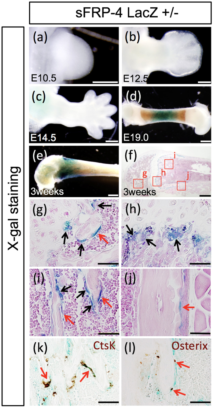
Figure 1. Temporally restricted manner of sFRP4 gene expression in the developing skeleton of mouse limb.
Whole-mount (a–e) and section views (f–l) of X-gal staining signals in sFRP4-LacZ knock-in reporter heterozygous mice (Supplementary Fig. 1. shows the sFRP4 gene targeting construct). (a–c) β-galactosidase activity is not detectable in the limb bud at early- and mid-embryonic stages. (d) Gradual β-galactosidase activity is detected around the neonatal stage. (g–j) Prominent X-gal stained signals in the mouse femur at 3 weeks of age. Strong β-galactosidase activity is detected at the mononucleic flat cells and the multinucleic cells in the epiphyseal nucleus (g), metaphysis (h), diaphysis (i) and periosteum (j). The mono-nucleic flat cells (red arrows) and multinucleic cells (black arrows) are seen in (g–j). (k,l) Immunohistostaining of X-gal stained sections of the diaphysial region of the femur with anti-Cathepsin Ab and anti-Osterix Ab confirms that β-galactosidase positive cells are localized in the osteoclastic and osteoblastic cell lineages (red arrows). Control experiments for X-gal staining are shown in Supplementary Fig. 2. Bars 500 μm (a–f), 50 μm (g–l).

