
Figure 4. Histological molecular characterization of sFRP4 LacZ/LacZ mice.
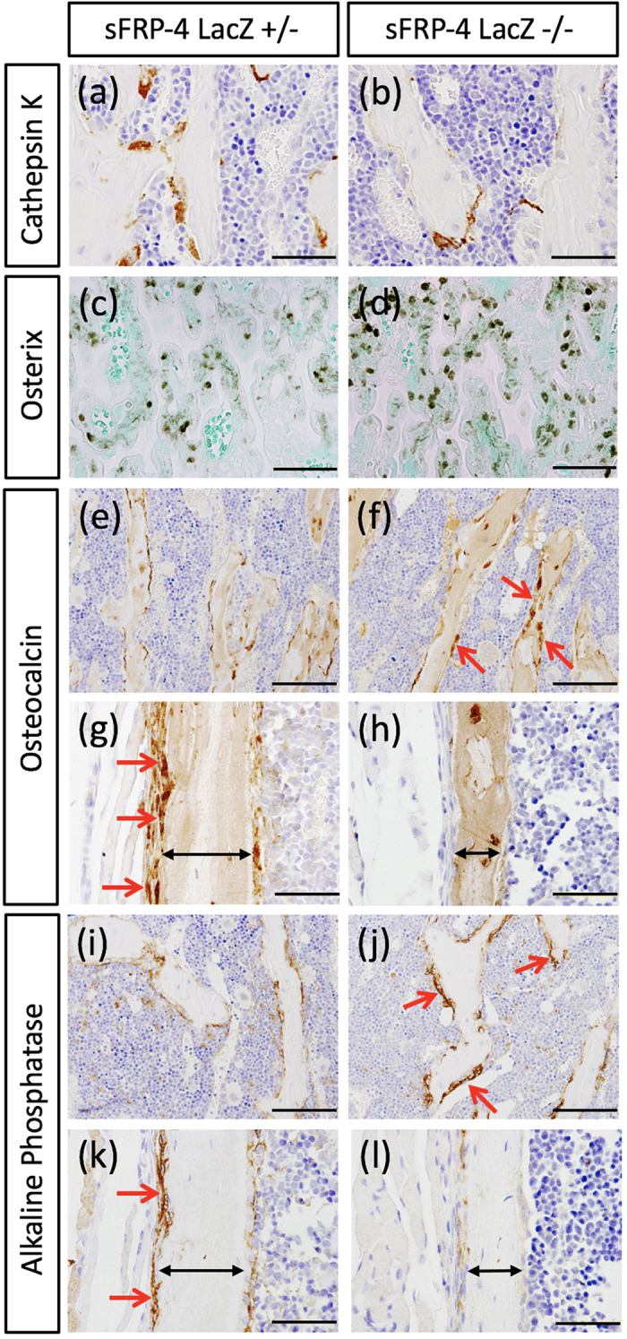
(a,b) A significant decrease of cathepsin K (osteoclast marker) protein expression is observed in sFRP4 LacZ/LacZ compared with that in sFRP4 LacZ/+ mice (positive stained cells ratio; control 31% and KO 10%). Supplementary Fig. 4. shows results of histochemical TRAP staining of monitored osteoclast activity. (c,d) Prominent Osterix expression in the trabecular region is observed in sFRP4 LacZ/LacZ compared with that in sFRP4 LacZ/+ mice (control 41% and KO 62%). (e–l) Expression pattern of bone deposition markers, osteocalcin and alkaline phosphatase: in the trabecular region, both markers are strongly expressed in sFRP4 LacZ/LacZ compared with those in sFRP4 LacZ/+ mice (osteocalcin; control 28% and KO 55%, alkaline phosphatase; control 30% and KO 53%); in the periosteal region, the expression of both markers is significantly repressed (g,h,k,l; red arrows, osteocalcin; control 39% and KO no-clear signal, alkaline phosphatase; control 79% and KO 16%). Bidirectional arrows denote periosteal width (g,h,k,l). Bars 50 μm (a,b,e,f,i,j), 100 μm (c,d,g,h,k,l).