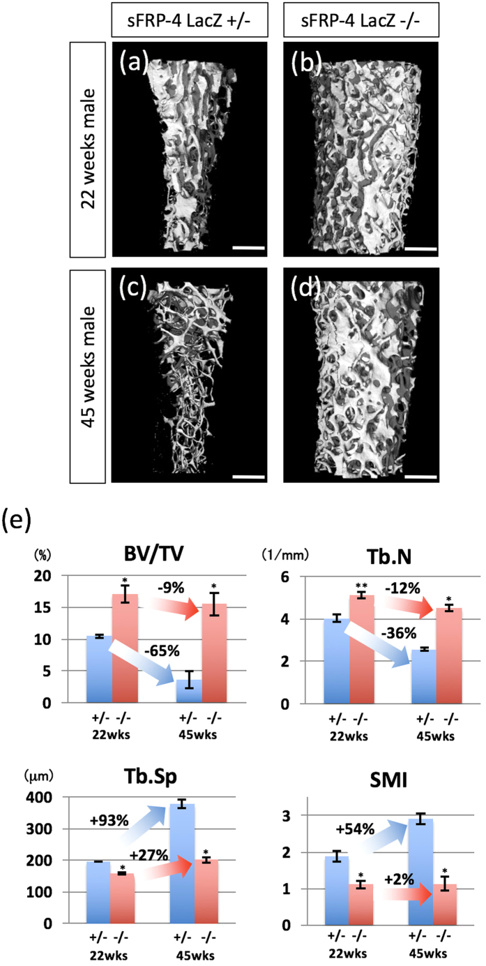
Figure 5. sFRP4 gene inactivation prevents age-related bone loss.
(a–d) Representative 3D μCT trabecular bone images of the distal femur of control and sFRP4 LacZ/LacZ mice at 22 and 45 weeks of age. In control mice, note greatly reduced trabecular mass indicative of trabecular osteopenia at 45 weeks (c) compared with that at 22 weeks (a). sFRP4 gene inactivation strikingly prevents age-related trabecular osteopenia in sFRP-4LacZ−/− (d) compared with control mice (c), at 45 weeks. (e) Quantification of Bone volume fraction (BV/TV), Trabecular number (Tb.N), Trabecular spacing (Tb.Sp) and Structure model index (SMI) of trabecular bone at distal femurs. Compared with control mice, sFRP4 LacZ/LacZ mice show no significant age-related differences (22 weeks vs. 45 weeks) in all structural bone parameters. The degree of variability (%) is shown in bar graphs. Error bars indicate the means ± SD (n=5); means ± SD in BV/TV: 10.44 ± 0.28 for 22wks+/−, 17.11 ± 1.32 for 22wks−/−, 3.6 ± 0.97 for 45wks+/− and 15.51 ± 1.8 for 45wks−/−, Tb.N: 4.03 ± 0.18 for 22wks+/−, 5.12 ± 0.16 for 22wks−/−, 2.57 ± 0.1 for 45wks+/− and 4.52 ± 0.14 for 45wks−/−, Tb.Sp: 196.37 ± 0.47 for 22wks+/−, 158.23 ± 2.1 for 22wks−/−, 378.97 ± 13.28 for 45wks+/− and 201 ± 7.78 for 45wks−/−, SMI: 1.88 ± 0.15 for 22wks+/−, 1.11 ± 0.11 for 22wks−/−, 2.91 ± 0.15 for 45wks+/− and 1.14 ± 0.2 for 45wks−/−. Statistical significance was determined by paired Student’s t-test. *P < 0.01 and **P < 0.05 versus sFRP4 LacZ +/−. Bars 900 μm (a–d).

