
Figure 2.
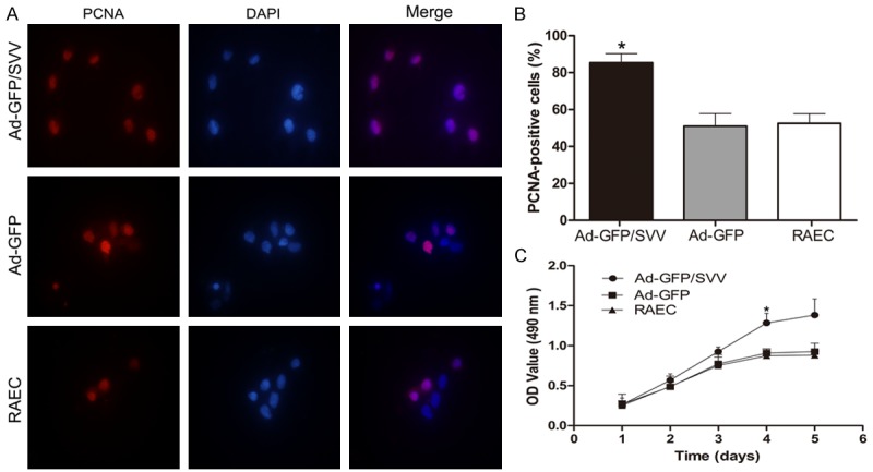
Effect of SVV on the proliferation of RAECs. A. Immunofluorescence staining for PCNA in RAECs after adenovirus transfection (original magnification: 1000×). B. Percentage of PCNA-positive cells after transfection. C. MTT assay. Proliferation of cells in Ad-GFP/SVV group significantly increased as compared to two control groups (Ad-GFP and RAECs, *P<0.05). SVV significantly enhanced the proliferation of RAECs. PCNA, proliferating cell nuclear antigen.