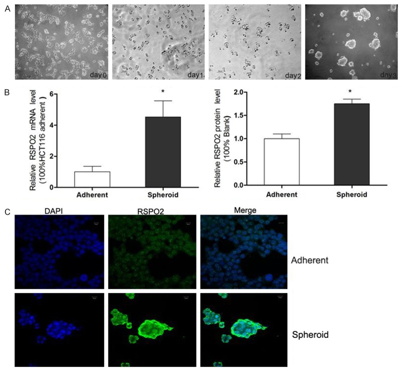
Figure 1.

RSPO2 expression in spheroid HCT116 cells. (A) Morphology of HCT116 adherent and spheroid cells. The HCT116 cells grew in medium containing FBS (serum added medium, SAM) was typical monolayer adherent cells (DAY 0), when the HCT116 cells subcultured in serum-free DMEM/F12 medium (SFM), they became suspended and turned into large spheres (DAY1-3) (×100). (B) RSPO2 mRNA expression and protein expression level in HCT116 adherent and spheroid cells, detected by qRT-PCR and western blotting. (C) Immunofluorescent staining of RSPO2 in HCT116 adherent (A) and spheroid cells (B). RSPO2 staining is green and nuclei are stained in blue.
