
Figure 4.
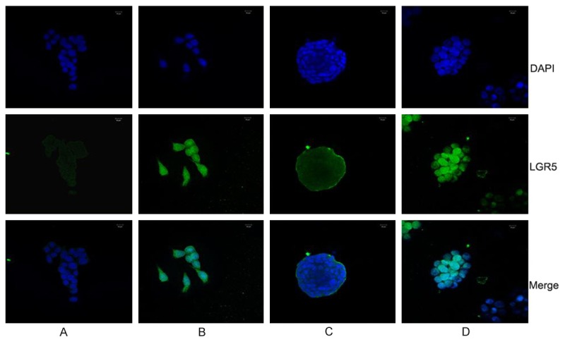
Immunofluorescent staining of LGR5 in HCT116 cells. A and B were HCT116 adherent cells with 0 ng/ml or 40 ng/ml recombinant RSPO2, C and D were HCT116 spheroid cells with 0 ng/ml or 40 ng/ml recombinant RSPO2; LGR5 staining is green and nuclei are stained in blue.