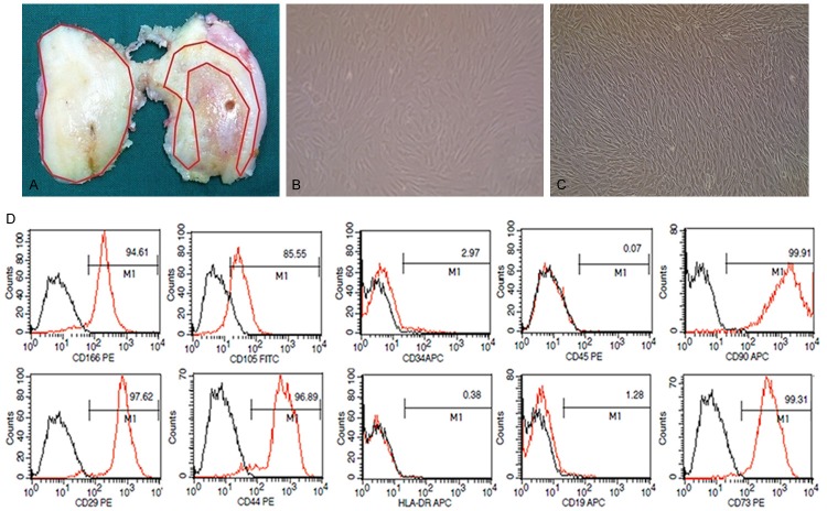
Figure 1.
Location of osteoarthritis cartilage specimen, cultivation and identification of cartilage-derived mesenchymal stem cells. A. Image of a representative distal femur of osteoarthritis patient who undergo a total joint replacement surgery. Locations used for the harvesting of cartilage are indicated by red lines: the normal cartilage sample is from healthy-appearing areas in lateral condyle, and the degraded cartilage sample is isolated from 0.5 to 1 cm areas around fibrosis or lesions in medial condyle. B. Chondrocytes on passage 1 are isolated from cartilage specimen (×100). C. CD105+/CD166+ cells on passage 2 are isolated by fluorescence-activated cell sorting (×100). D. Cell surface antigen expression of selected markers on CD105+/CD166+ cells, which met the immune-phenotype criteria of MSC.

