
Figure 3.
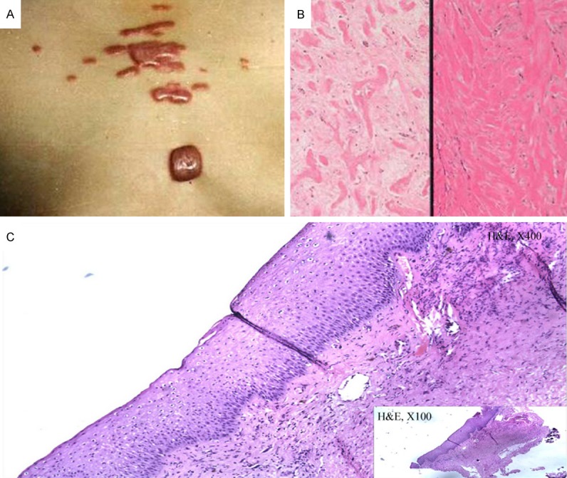
Photograph of one sporadic case and Biopsy of one patient and one control sample. A. This picture shows the chest of one of the sporadic cases. B. Biopsy of a patient revealed a typical fibroplasia in the corium layer. C. Biopsy of one control sample shows the normal corium layer.