
Figure 1.
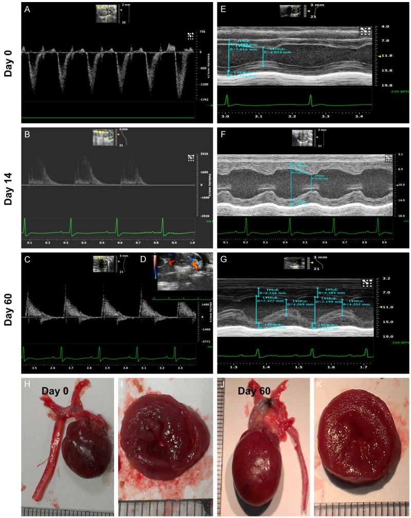
Echocardiography and pathological findings provided evidence of pathological hypertrophic cardiomyopathy (PHC). (A-C) Illustrating the transthoracic echo Doppler findings of velocity of blood flow in ascending aorta of DPP-4 deficiency rats at baseline and by days 14 and 60 after transverse aortic constriction (TAC) procedure. The results showed that the velocity was notably increased at day 14 and more remarkably increased by day 60 after TAC procedure as compared with the baseline level. The systolic blood flow velocity of ascending aorta at baseline, and days 14 and 60 after TAC, was < 100 mm/s, 3500 mm/s and 4200 mm/s, respectively. (D) The transthoracic echocardiography showed remarkably mosaic turbulence blood flow in ascending aorta by day 60 after TAC procedure. (E-G) M-mode showing the thickness of interventricular septum (IVC) and posterior wall of left ventricle were notably increased by day 14 and further remarkably increased by day 60 after TAC induction as compared with baseline level in the DPP-deficiency rat. (H and J) The anatomical finding showing the appearance of whole heart size in DPP-deficiency rat was remarkably larger by day 60 after TAC induction as compared with animal without receiving TAC procedure. (I and K) The pathological finding (i.e., cross section picture at papillary muscle level) showed the notably concentric hypertrophy of LV muscle at day 60 (K) as compared with day 0 (I). Data from (A-K) suggesting the TAC model for inducing PHC was successfully created.