
Figure 2.
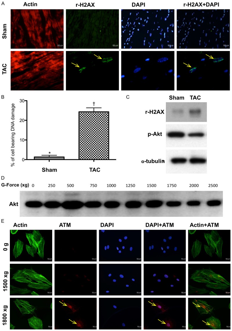
The results of pilot experimental and in vitro studies for proving pressure overload activated DNA damage signaling (Figures 1, 2 and 3) (n = 6). A. Immunofluorescent (IF) microscopic (200x) finding for identification of γ-H2AX+ cells (yellow arrows) in sham controls and in animals after day-60 transverse aortic constriction (TAC) procedure. B. Statistical results showing the percentage of cell bearing DNA damage was significantly higher in TAC group than in sham control, * vs. †, p < 0.0001. C. By day 60 after TAC procedure, the protein expression of γ-H2AX was notably increased whereas the protein expression of phosphorylated (p)-Akt was remarkably reduced in TAC animals than in sham-control animals. D. In vitro study showing that the protein expressions of Akt was notably inhibited after the gravity (G) force was increased up to ≥ 1500. E. The number of ATM+ cellular expression (yellow arrows) was notably stepwise increase after increased G force application to the H9C2 cells.