
Figure 3.
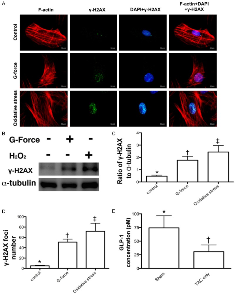
Compared the protein and cellular expressions of γ-H2AX after the G force (2500 xg) application and 30-minute H2O2 (200 µM) treatment (n = 6) and determined the circulating GLP-1 level by day 60 after TAC procedure (n = 4). A. Confocal microscopic (400x) finding for the expressions of γ-H2AX+ cells in situations of control, G force, and H2O2 treatment, respectively. B. The protein expression of γ-H2AX was remarkably higher in G force treatment and more remarkably higher in H2O2 treatment as compared with the control. C. Analytical result of the ratio of γ-H2AX to α-tubulin, * vs. other groups with different symbols (*, †, ‡), p < 0.001. D. Analytical result of counted γ-H2AX foci formation (green color), * vs. other groups with different symbols (*, †, ‡), p < 0.0001. E. Analytical result of day-60 circulating glucagon-like peptide (GLP)-1 level, * vs. †, p = 0.047. All statistical analyses were performed by one-way ANOVA, followed by Bonferroni multiple comparison post hoc test. Symbols (*, †, ‡) indicate significance (at 0.05 level).