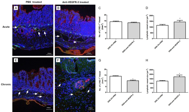
Figure 6.
Changes in lymphatic vessels after VEGFR-3 blocking in acute and chronic colitis. Double immunofluorescence staining was performed for differentiating lymphatic vessels (green, CD31+LYVE-1+) from blood vessels (red, CD31+LYVE-1-), with DAPI staining for nucleus (blue). Compared with PBS-treated mice (A), mice with anti-VEGFR-3 antibody treatment had no significant change in LVD (B, C, P > 0.05), but significantly enlarged lymphatics were observed (D, t test, P < 0.05) in acute colitis mice. In chronic colitis mice, LVD was significantly decreased in anti-VEGFR-3-treated mice, as compared to PBS-treated mice (E-G, t test, P< 0.05), accompanied by dramatically enlarged and distorted lymphatics (F, H, t test, P < 0.05).

