
Figure 3.
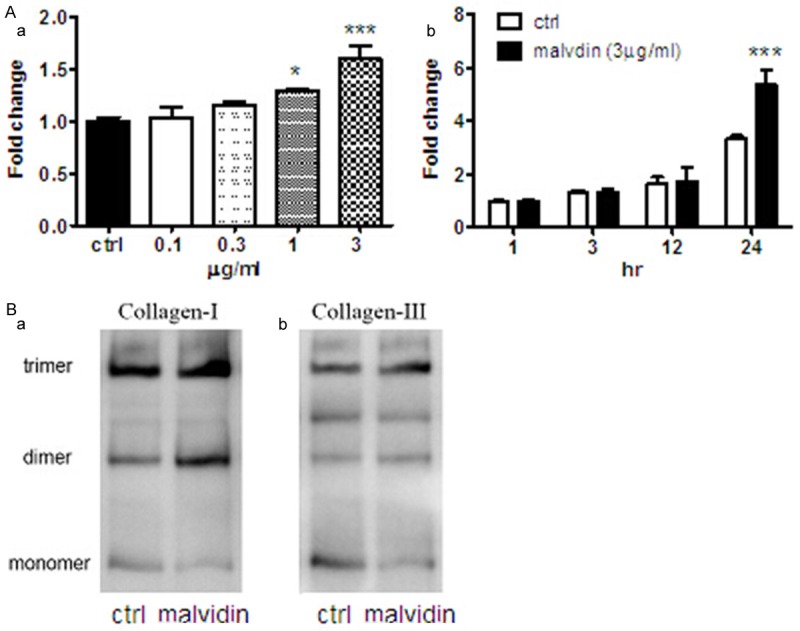
Anthocyanins stimulated collagen expression and secretion in cardiac fibroblast cells. A. (a) After treatment with anthocyanins for 24-h, the extracellular medium of fibroblast was collected and concentrated for measurement of collagen content by Picrosirius Red staining. The results are presented as fold change against the control value. (b) The extracellular fibrillar collagen was measured after treatment with 3 ug/ml anthocyanins for various time points. The results are presented as fold change against the control value at 1-h. ***P < 0.001; *P < 0.05 vs corresponding ctrl, n=6. B. Protein samples derived from fibroblast cells following treatment with anthocyanin for 24-h were subjected to Western blot analysis and probed with rabbit anti-type I collagen antibody (a) and rabbit anti-type III antibody (b). Data shown are representative results from three independent experiments.