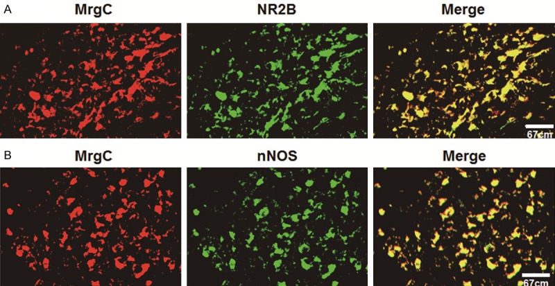
Figure 5.

Confocal images showing the localization of (A) NR2B-IR or (B) nNOS-IR with MrgC-IR in SCDH neurons. (A) NR2B-IR neurons are identified by Alexa Fluor 488 fluorescence (green), whereas MrgC-IR-positive neurons by Alexa Fluor 594 (red). Co-localization of NR2B-IR with MrgC-IR in DRG neurons is identified by yellow. (B) nNOS-IR neurons are identified by Alexa Fluor 488 fluorescence (green) whereas MrgC-IR-positive neurons by Alexa Fluor 594 (red). Co-localization of nNOS-IR with MrgC-IR in DRG neurons is identified by yellow. Scale bar = 50 μm.
