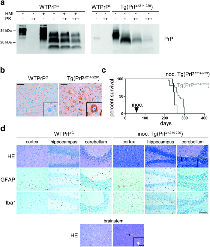
Figure 6. PrPΔ214–229 is partially PK resistant and accelerates disease in syngeneic host.
(a) Western blot analyses of PrP in brain homogenates after PK digestion. 4 μl of 10% total brain homogenate were treated with increasing amounts of PK (10 μg/ml (+); 20 μg/ml (++) and 40 μg/ml (+++)). A wild type prion diseased mouse (inoculated with RML prions) was used as a positive control. WTPrPC was detected with the POM1 antibody whereas Tg(PrPΔ214–229) was detected with the 3F4 antibody and therefore only unspecific bands are observed for WTPrPC in this blot. Note that Tg(PrPΔ214–229) is PK resistant at the concentration widely used to define PrPSc (20 μg/ml), but compared to the prion diseased mice brain is less resistant at higher concentrations of PK. (b) Representative immunohistochemistry of cortical mouse brain sections stained with SAF84 antibody after PK digestion. Higher magnification shows accumulation of PK-resistant PrP surrounding the nucleus in Tg(PrPΔ214–229). Scale bar in (b,d) is 100 μm. (c) Kaplan-Meier survival curve of Tg(PrPΔ214–229) (grey) and of Tg(PrPΔ214–229) inoculated with brain homogenates obtained from clinically diseased Tg(PrPΔ214–229) mice (black). A significant acceleration of the disease is observed (Breslow test: p* = 0.007). (d) Brains from Tg(PrPΔ214–229) mice inoculated with Tg(PrPΔ214–229) brain homogenates (inoc. Tg(PrPΔ214–229)) presented very faint spongiosis (as seen with HE staining) mainly in cerebellum and striatum and no overt astro-/microgliosis (as seen with immunohistochemistry for GFAP and Iba1). Scale bar in the magnified picture is 25 μm.

