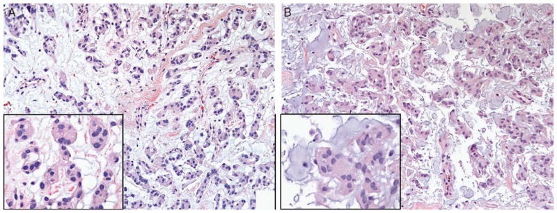
FIGURE 2.
A, Lobulated tumor cells with vacuolated cytoplasm and well-demarcated cell borders characteristic of chordoid meningioma (inset) arising in a delicate fibrous background. B, Chordoid meningioma can also exhibit nested tumor cells (inset) with a more myxoid background. When unaccompanied by areas of typical meningioma or in a diminutive biopsy sample, correct identification of this meningioma variant can be challenging.

