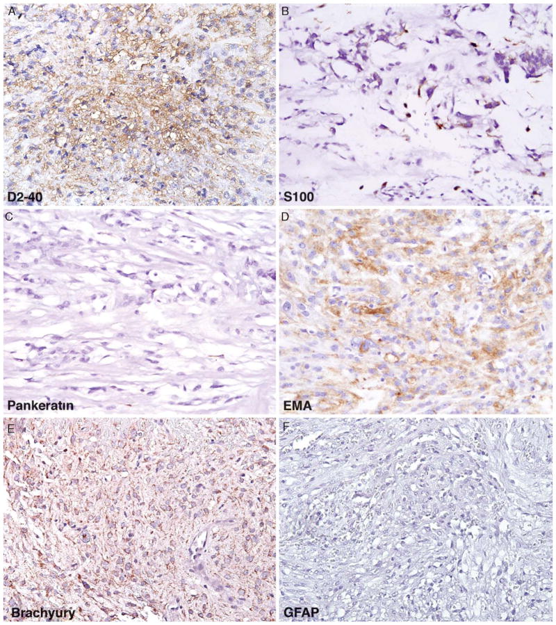
FIGURE 4.
Typical immunoprofile of chordoid meningioma on the basis of the results of this study include (A) consistent membranous to cytoplasmic positivity with D2-40, (B) patchy S100 positivity, (C) predominantly negative staining with pankeratin, (D) strong cytoplasmic positivity for EMA, (E) background cytoplasmic staining (non-nuclear) with brachyury, and (F) negative staining with GFAP. EMA indicates epithelial membrane antigen; GFAP, glial fibrillary acidic protein.

