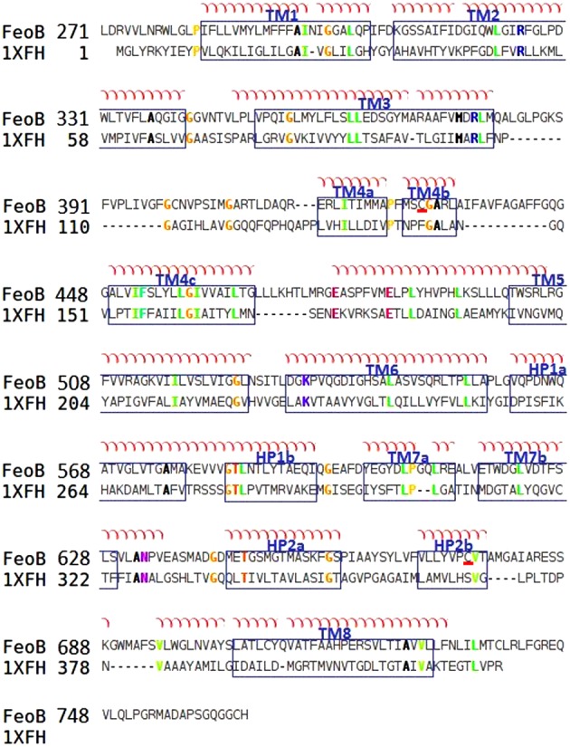
Figure 4. Alignment between the TM domains of FeoB from P. aeruginosa (FeoB) and of the glutamate transporter from P. horikoshii (1XFH).

The TM segments of FeoB are arranged according to the crystal structure of the glutamate transporter and are visualized in blue boxes. The alignment was made using ClustalW and adjusted manually. Conserved cysteine residues are underlined in red.
