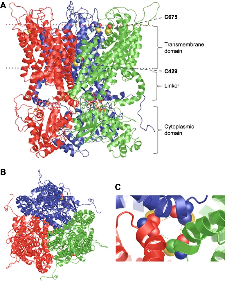
Figure 5. Homology model of FeoB from P. aeruginosa.
The three subunits are coloured green, red and blue. The conserved cysteine residues from TMH4 and TMH7 are drawn using space-filled atoms. The GTP ligands are in stick representation. The FeoB homotrimer is viewed either from the plane of the membrane (A) or and from the extracellular side of the membrane (B). TMH4s from the three monomers form a central pore. (C) Close-up view of the pore-forming TMH4s from the extracellular side of the membrane. The distances between the α-carbons of the TMH4 cysteine residues, indicated by orange dashed lines, are 8.5 Å.

