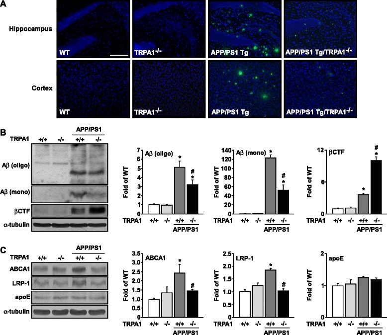
Fig. 3.

Ablation of TRPA1 channel function decreases Aβ deposition in brain lesions of APP/PS1 Tg mice. (a) Immunostaining of specimens of hippocampus and cortex from 8-month-old WT, TRPA1−/−, APP/PS1 Tg and APP/PS1 Tg/TRPA1−/− mice with anti-Aβ antibody, then FITC-conjugated secondary antibody. (b, c) Western blot analysis of protein levels of oligomer (oligo) or monomer (mono) Aβ, βCTF, ABCA1, LRP-1, apoE and α-tubulin in brain specimens. Bar = 100 μm. Data are mean ± SEM from 8 mice in each group. *, P < 0.05 vs. WT mice. #, P < 0.05 vs. APP/PS1 Tg mice
