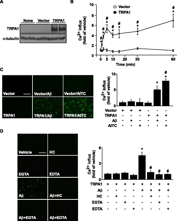
Fig. 6.

The essential role of TRPA1 in Aβ-induced calcium influx in HEK293 cells. (a) Western blot analysis of TRPA1 and α-tubulin protein levels in non-treated, vector- or TRPA1-transfected HEK293 cells. (b) Ca2+ influx in HEK293 cells transfected with vector or TRPA1 plasmid, then treated with Aβ (2 μM) for 0–60 min. Fluo-8 calcium assay of the intracellular level of Ca2+ in (c) HEK293 cells transfected with vector or TRPA1 plasmid for 24 h, then treated with Aβ (2 μM) or AITC (10 μM, a TRPA1 agonist) for 5 min and (d) after transfection with TRPA1 plasmid, HEK293 cells pretreated with a TRPA1 antagonist HC030031 (HC) 10 μM, EGTA 0.5 mM and EDTA 0.5 mM for 2 h, then with Aβ for 5 min. Fluorescence images were photographed by fluorescence microscopy. Bar = 100 μm. Data are mean ± SEM from 5 independent experiments. *, P < 0.05 vs. 0 min or TRPA1 transfection alone. #, P < 0.05 vs. vector-transfection alone or TRPA1 transfection + Aβ treatment
