Fig. 1.

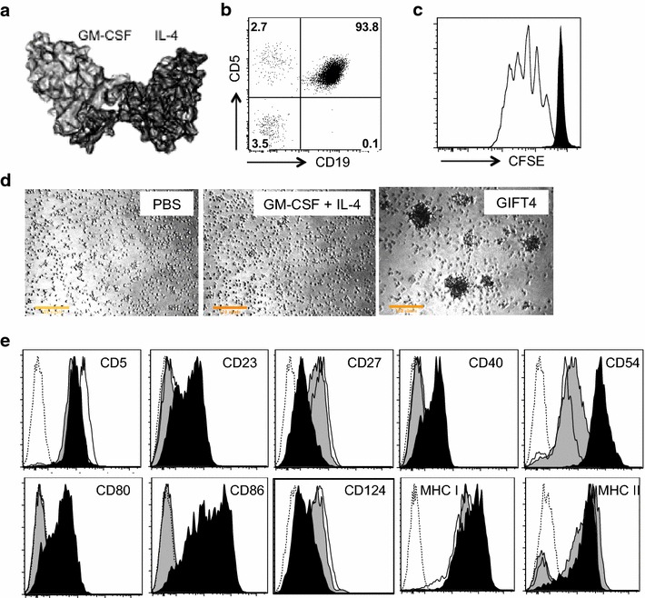
Phenotype of GIFT4-CLL cells. a Predicted 3D structure of GIFT4 protein. b A representative of CD19+CD5+ primary CLL cells in PBMC of subjects. c Purified normal human B cells (White) or CLL cells were labeled with CFSE dye and treated with GIFT4 protein for 5 days; cell proliferation was analyzed by FACS. d CLL cells treated with GIFT4, GM-CSF and IL-4 or PBS were photographed under a microscope (×10). e CLL cells treated with GIFT4 (dark), GM-CSF and IL4 (gray), or PBS (white) were subjected to FACS analysis with a panel of antibodies against B cell markers or with antibody isotype control (Dash). Data are representative of one of four repeated experiments using samples from subjects No. 1, 2, 3 and 4
