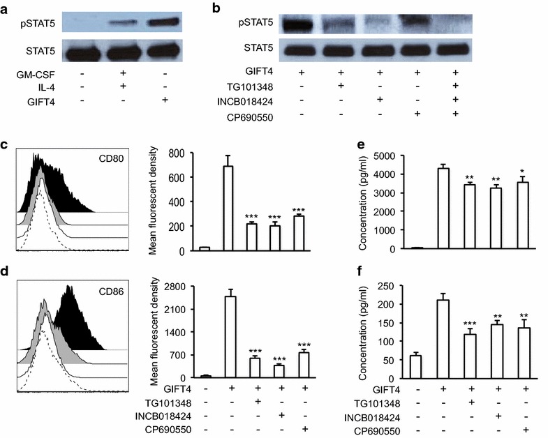
Fig. 3.

GIFT4-induced STAT5/JAK signaling in CLL cells. a Primary CLL cells were stimulated with GIFT4, GM-CSF and IL-4 or PBS for 20 min. The cells were harvested and lysed. Ten microgram of proteins in the cell lysate was subjected to Western blot analysis with anti-pSTAT5 or anti-STAT5 antibodies. b CLL cells were treated with GIFT4 protein in presence or absence of inhibitors for JAK2 (TG101348), JAK1/2 (INCB018424) or JAK3 (CP690550). The cell lysate was subject to Western blot analysis with anti-pSTAT5 and anti- STAT5 antibodies. c, d CLL cells were stimulated with GIFT4 protein for 5 days, supplemented with JAK inhibitors. CD80 (c) and CD86 (d) expression on CLL cells treated with GIFT4 protein (Dark), or GIFT4 with JAK2 inhibitor (Dash), JAK1/2 inhibitor (White), JAK3 inhibitor (Gray) was analyzed by FACS. Mean fluorescent density was also quantified. e and f Cytokine IL-2 and IL-6 secretion by the treated CLL cells was quantified by ELISA with anti-IL-2 and anti-IL-6 antibodies, and calculated from three repeated experiments using samples from subjects No. 6, 8 and 9
