
Fig. 1.
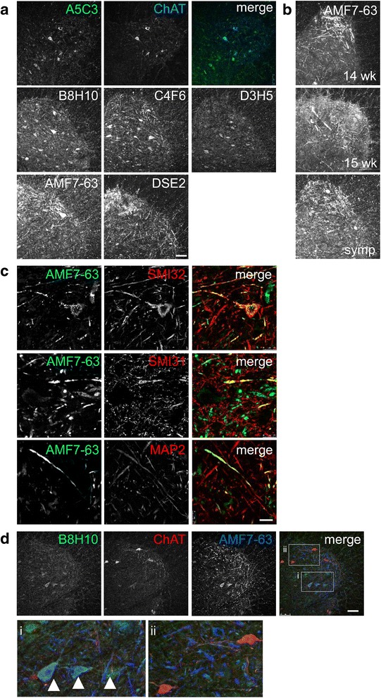
Misfolded SOD1-specific antibodies have distinct labeling patterns in SOD1G93A rat spinal cords. Immunohistochemistry for misfolded SOD1 in SOD1G93A lumbar spinal cords. a Lumbar sections of a symptomatic SOD1G93A rat were stained with misfolded SOD1 specific antibody A5C3 (green) and co-labeled with ChAT (blue). Additional representative images of B8H10, C4F6, D3H5, DSE2-3H1 and AMF7-63 are also shown. b The AMF7-63 antibody detects fibrils in pre-symptomatic, 14 and 15 week SOD1G93A rat spinal cords. c Symptomatic SOD1G93A rat spinal cord was labeled with AMF7-63 (green) and SMI32 (red), SMI31 (red), or MAP2 (red). d Lumbar sections labeled with misfolded SOD1 antibodies AMF7-63 (blue), B8H10 (green), and co-labeled with ChAT (red). Two to three animals of each genotype were analyzed. Scale bar = 100 μm for (a, b and d) and 25 μm for (c)