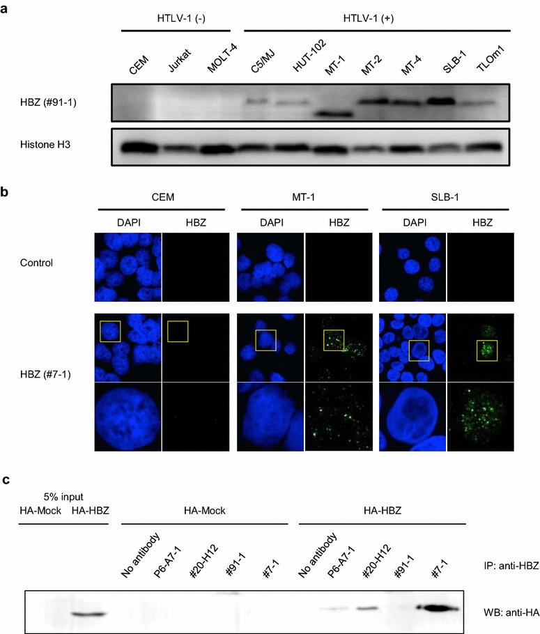
Fig. 2.

HBZ protein expression in HTLV-1-infected cell lines. a HBZ protein expression in HTLV-1-infected cell lines was analyzed by western blotting using anti-HBZ mAb #91-1 (raised against peptide #3, rat IgG1), which gave highest sensitivity among four mAbs (i.e. P6-A7, #20-H12, #91-1, #7-1). Histone H3 was used as a loading control for the nuclear fraction. b HBZ protein expression in HTLV-1-infected cell lines (MT-1 and SLB1) was analyzed by immunofluorescence microscopy using anti-HBZ mAb #7-1 (raised against recombinant HBZ, mouse IgG2b), which gave highest sensitivity among four mAbs (i.e. P6-A7, #20-H12, #91-1, #7-1). Three independent experiments were performed, and representative data are shown in each figure. c Immunoprecipitation assay using four different anti-HBZ mAbs. P6-A7, #20-H12, and #7-1 were available for immunoprecipitation
