
Fig. 4.
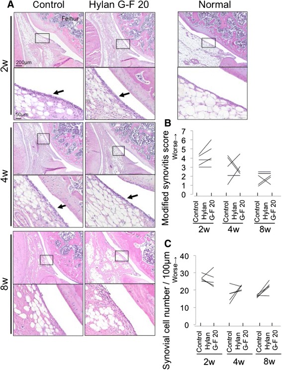
Analyses of histological observations for the infrapatellar fat pad. a Sagittal sections stained with hematoxylin and eosin. An 18-week-old rat was used as a normal. Boxed areas in the upper panels are shown at a higher-magnification view in the lower panels. Arrow indicates increased number of synovial cells. b Modified synovitis score for infrapatellar fat pad. c Synovial cell number/100 μm synovium