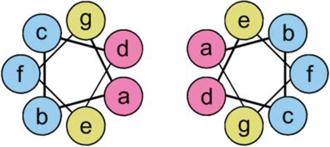
Fig. 25.
A schematic of a coiled–coiled dimer containing the (abcdefg) motif. The ‘a’ and ‘d’ residues are typically hydrophobic amino acids, while the ‘e’ and ‘g’ residues are charged moieties. The dashed lines represent hydrophobic and charge interactions resulting in the coiled-coil structural motif.
Obtained and modified from Jonker et al. [415].

