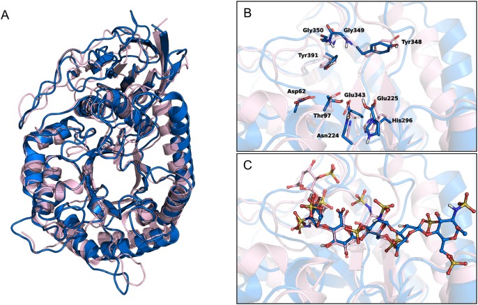
Fig. 7.
(A) Superposition of human heparanase crystal structure (pink ribbons, PDB code: 5E9C) and the homology model of GS3 construct (blue ribbons). (B and C) Close-up views of the ligand-binding site of heparanase crystal in complex with ligand dp4 (ΔHexA2S-GlcNS6S-IdoA-GlcNS6S, pink carbons) and heparanase GS3 construct in complex with fondaparinux (blue carbons). (B) depicts the residues delimiting the catalytic sites, (C) shows the ligand molecules dp4 and roneparstat.

