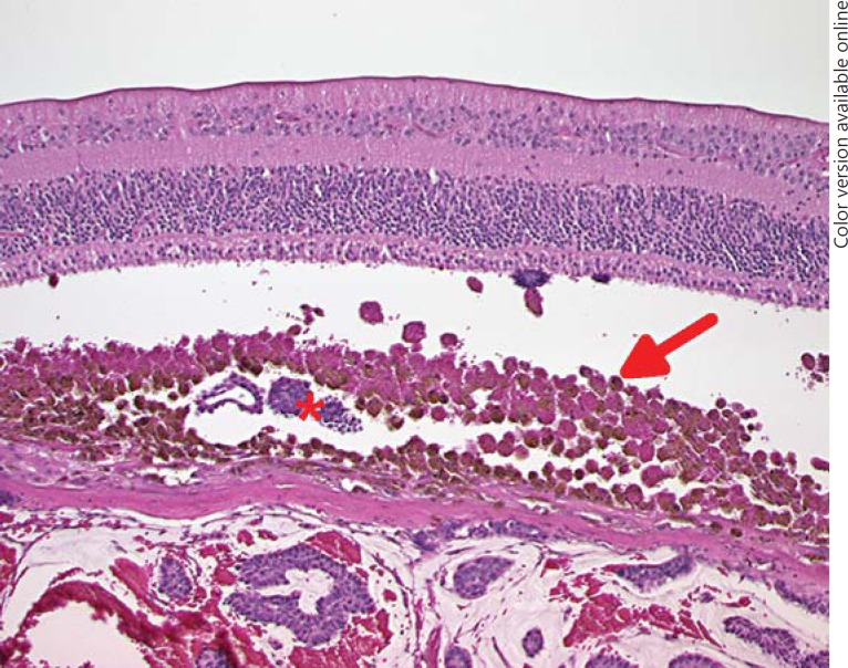
Fig. 4.
An accumulation of macrophages is present beneath the retina (arrow). These cells are packed with lipofuscin and pigment. Tumour cell sheets are present in the underlying choroid as well as in the sub-retinal space (asterisk), indicating an invasion of the Bruch's membrane elsewhere. Photoreceptor degeneration and RPE layer disruption are also present. HE. ×100.

