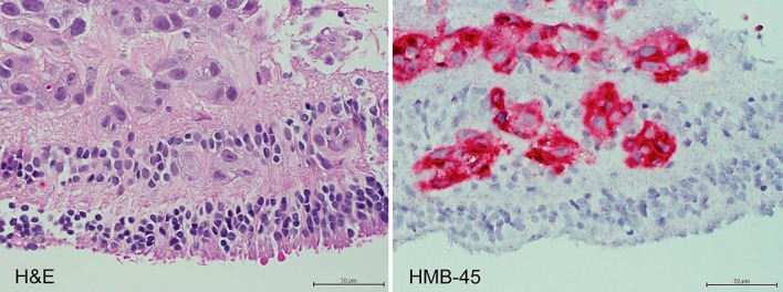
Fig. 4.

Histopathology of retinal biopsy from the affected eye. Left: amelanotic, dis-cohesive malignant cells with enlarged nuclei are scattered throughout the retina, most prominently in the inner layers (×400, HE). Right: malignant cells are replacing the inner retina (×400, HMB-45).
