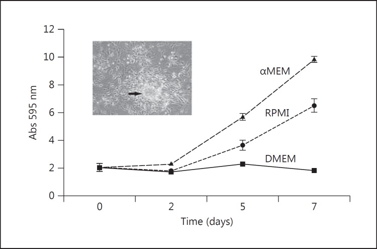
Fig. 1.
Growth rate of isolated primary uveal melanoma cells from a monosomy 3 patient in different media. The cells were grown in either RPMI + 10% FCS, DMEM + 10% FCS, or 1:1 αMEM: Quantum 3-21 + 10% FCS. The cells grew more efficiently in the latter medium and demonstrated the formation of small clumps of cells that could be detached into the medium.

