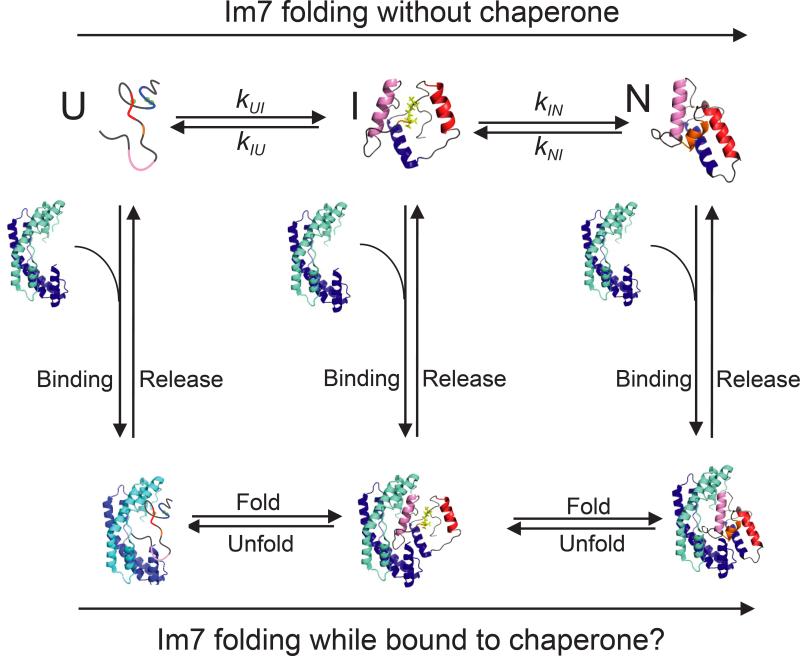
Figure 1.
Folding of the protein Im7 in the presence and absence of the chaperone Spy. Im7 is shown as a multicolored protein that is helical in both the folding intermediate (I) and in the native folded state (N) and lacks any persistent secondary structure in the unfolded state (U). Spy is shown as a blue cradle-shaped homodimer. Spy binding and release are shown as vertical arrows.

