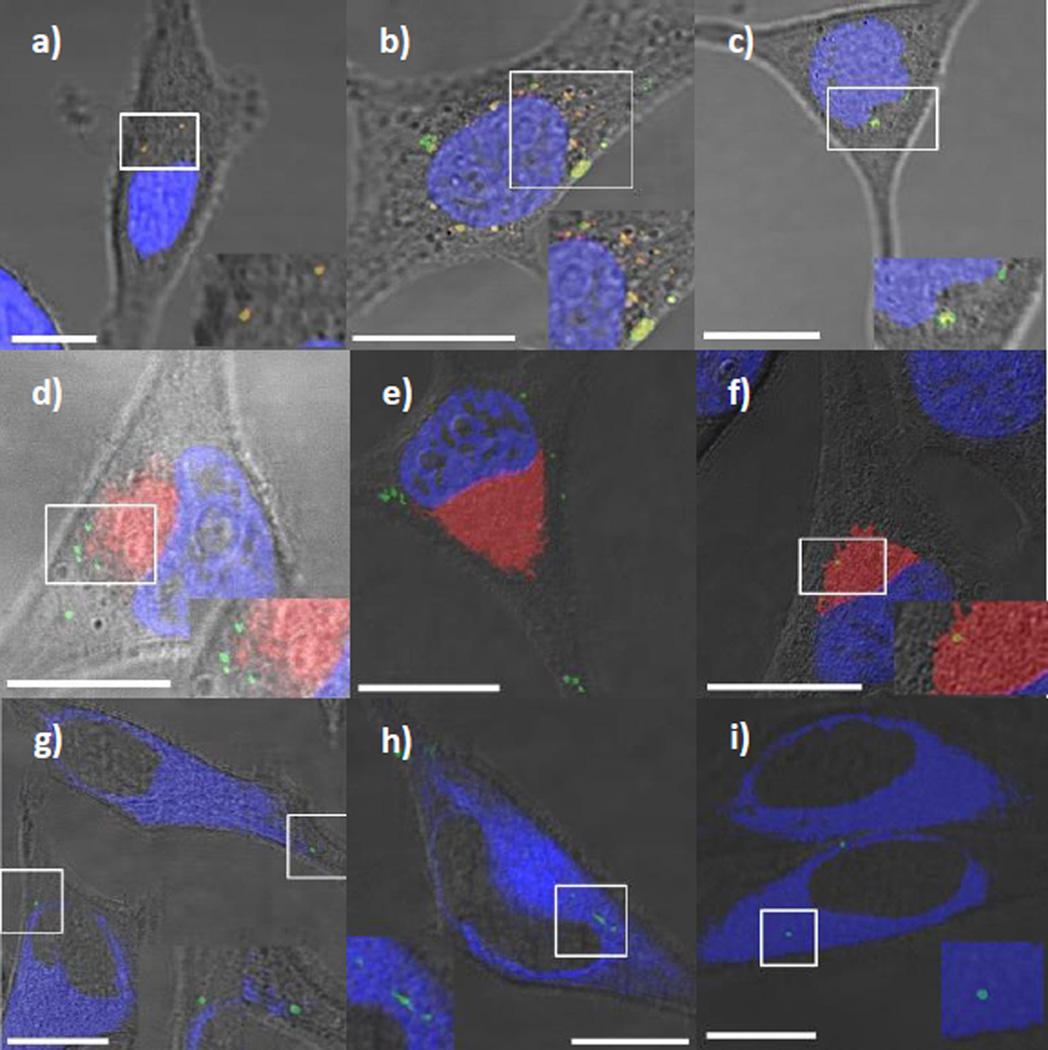
Fig. 3.
LSCM images of HeLa cells inoculated with FMSNs (a,d,g), PEGC-FMSNs (b,e,h) and CTxB-FMSNs (c,f,i) in the presence of Lysotracker Red (DND-99) (red) (a–c), BODIPY-Ceramide TR conjugated to BSA™ (red) (d–f) and ER-Tracker™ Blue-White DPX (blue) (g–i). All FMSN materials are shown as green spots. The yellow spots result from the colocalization of FMSN materials with either Lysotracker Red (DND-99) or BODIPY-Ceramide TR conjugated to BSA™. The white spots resulted from the colocalization of FMSN materials with ER-Tracker™ Blue-White DPX. Insets on the bottom right corner (left corner for Fig. 3h) of the micrographs show a close-up of the highlighted region in the white square. All the scale bars are 10 µm in size.

