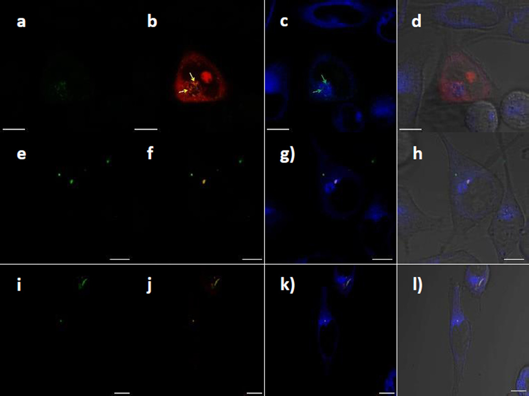
Fig. 5.
LSCM images of HeLa cells inoculated with PI-loaded CTxB-FMSNs, FMSNs and PEGC-FMSNs. (a,e,i) CTxB-FMSNs, FMSNs and PEGC-FMSNs (green). (b,f,j) CTxB-FMSNs, FMSNs or PEGC-FMSNs super-imposed with PI molecules (red); yellow arrows in Figure 5b indicate the colocalization of CTxB-FMSNs and PI molecules. (c,g,k) ER-stained images super-imposed with CTxB-FMSNs, FMSNs or PEGC-FMSNs; white arrows in Figure 5c indicate the colocalization of CTxB-FMSNs and the ER organelle. (d,h,l) Super-imposed micrographs of the previous images with the DIC channel. All the scale bars are 10 µm in size.

