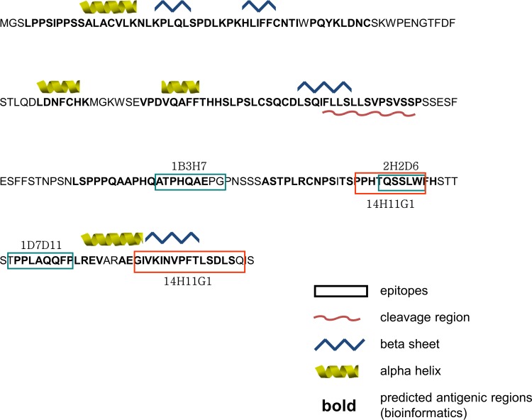
Fig 2. Gag-H protein sequence, structure and antibody epitopes.
Protein features, predicted using Geneious 7.1.4 [37], are indicted as follows: alpha helix (yellow helix), beta sheet (blue zigzag), signal cleavage region (red wavy line) and antigenic region (bold). Epitopes were determined using a sequential overlapping peptide library. The minimal epitopes defined as the amino acid sequence shared by all peptides detected by the antibody are labeled in boxes and the names of monoclonal antibodies are indicated.

