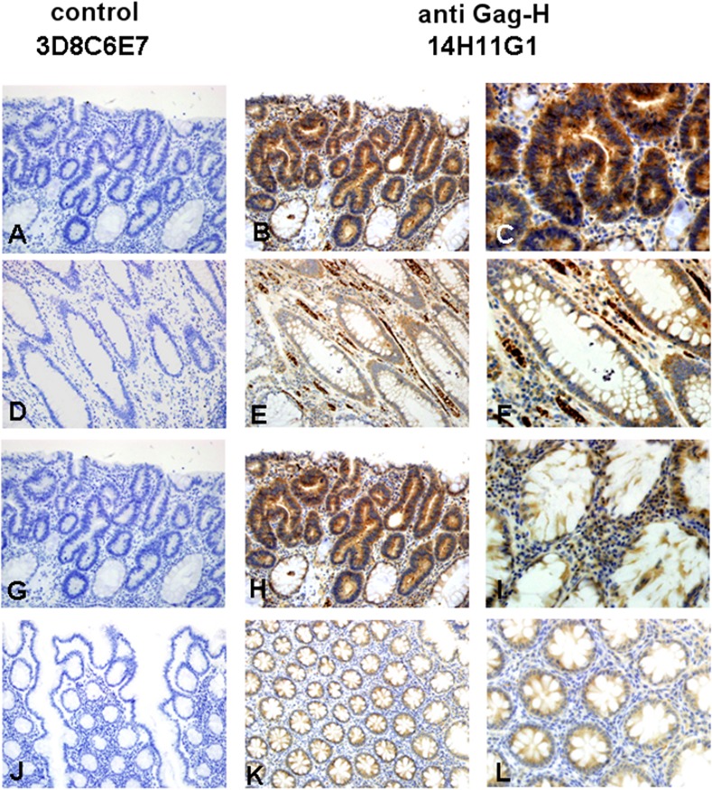
Fig 7. Examples of Gag-H immunohistochemistry in two representative clinical MSI cases.

The upper row represents the tumor and the bottom row the normal tissue of the cases. Both cases: No. 8 (A-F) and No. 11 (G-L) showed strong cytoplasmic immunostaining of Gag-H in tumor and in normal tissue (B, E, H, K by x20 objective; C, F, I, L by x40 objective). Negative immunostaining was achieved with the control antibody 3D8C6E7 (A, D, G, and J by x20 objective).
