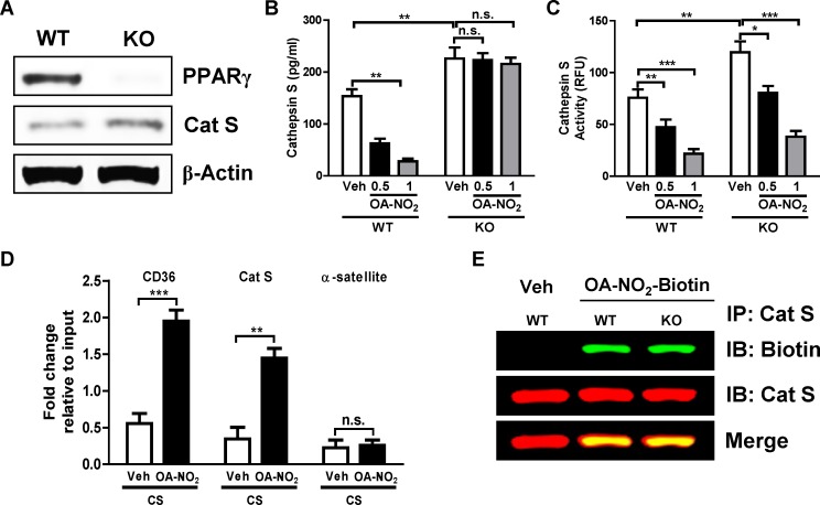
Fig 2. NFAs’ inhibition of Cat S expression but not activity, is PPARγ-dependent.
(A) Western blots for PPARγ and Cat S in whole cell extracts from AMs isolated from BAL fluid of C57BL/6 (WT) and Tie2 Cre-PPARγflox/flox (KO) mice. (B, C) Mice were exposed to cigarette smoke (CS) for 14 days, AMs were isolated and then treated with 0.5 and 1 μM OA-NO2 or Veh for 6 h. (B) Cat S expression and (C) activity in culture media. (D) Following AM isolation and OA-NO2 (1 μM) treatment as above, chromatin was crosslinked and immunoprecipitated with PPARγ antibody; the antibody-bound DNA-protein complexes were then subjected to real-time PCR with primers specific for PPRE sites in Cat S and CD36 promoter regions, α-satellite was used as control. (E) As above, AMs were collected from CS-exposed mice and treated with 5 μM biotin-labeled OA-NO2 or Veh for 6 h. Total protein extracts were prepared, Cat S was immunoprecipitated and Western blotting was performed with anti-biotin and -Cat S antibodies. Data are representative of two to three independent experiments with cells obtained from n = 14–18 mice/group (cells from 3 mice pooled together). *P < 0.05, **P < 0.01, ***P < 0.001, n.s. = non-significant.

