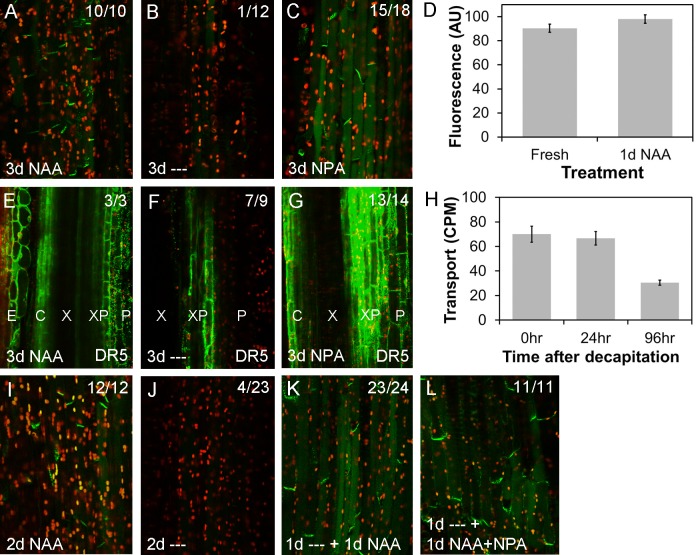
Fig 5. Auxin promotes PIN1 localization at the plasma membrane.
For confocal images (A–C; E–G, I–L), green signal indicates GFP, red signal is chloroplast autofluorescence. Images are from hand-cut sections of ~2 cm basal inflorescence internode segments from of 6-wk-old plants, held vertically between agar blocks for the times indicated and imaged at the apical end of the segment. A–C) PIN1-GFP expression in xylem parenchyma cells of PIN1pro:PIN1-GFP segments treated apically with 1 μM NAA for 3 d (A), no treatment (‘—’) for 3 d, (B) or 1 μM NPA for 3 d (C). Numbers in the right hand corner indicate the number of segments/the number examined in which basal, polar PIN1 localization was seen in this treatment. D) Quantification of PIN1-GFP levels (in arbitrary units, AU) at the basal plasma membrane in freshly harvested PIN1pro:PIN1-GFP stem segments versus segments treated apically for 1 d with 1 μM NAA. Forty-five membranes were analyzed per treatment (five each of nine individual stem segments); bars represent s.e.m. E–G) GFP expression in DR5rev-GFP stem segments treated apically with 1 μM NAA for 3 d (E), no treatment (‘—’) for 3 d, (F) or 1 μM NPA for 3 d (G). Numbers in the right-hand corner indicate the number of segments/the number examined in which the pattern shown was observed. H) The ability of basal internode segments 0, 24, and 96 hours after excision from the plant to transport radio-labelled IAA to their basal 5 mm (measured as counts per minute, CPM) during a 6 hour incubation with their apical ends immersed in radio-labelled auxin. The segments were held vertically between agar blocks for 0, 24, and 96 hours, as in A–G, before transfer to Eppendorf tubes for the auxin transport assay. n = 14–16; bars indicate s.e.m. I–L) PIN1-GFP expression in xylem parenchyma cells of PIN1pro:PIN1-GFP segments treated apically with 1 μM NAA for 2 d (I), no treatment (‘—’‘) for 2 d, (J) or no treatment for 1 d followed by 1 μM NAA for 1 d (K), or 1 μM NAA and 1 μM NPA for 1 d (L). Numbers in the right hand corner indicate the number of segments/the number examined in which basal, polar PIN1 localization was seen in this treatment.

