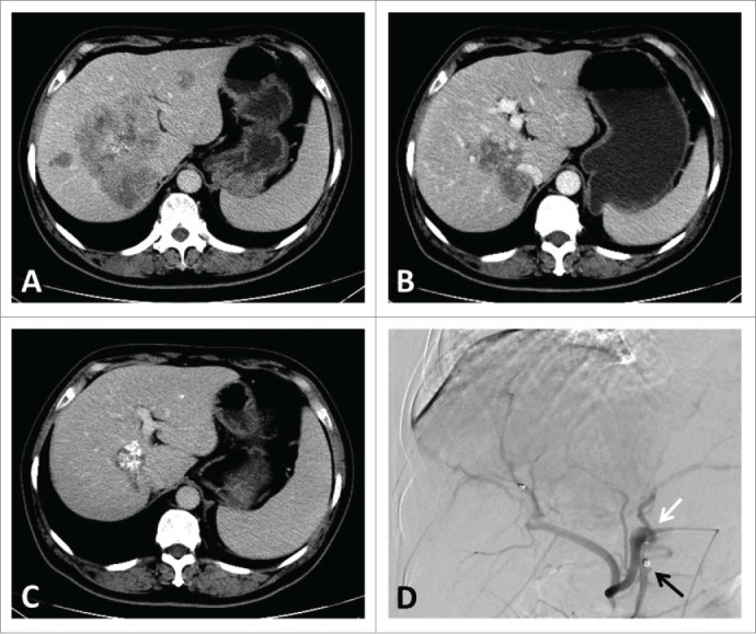
Figure 1.

A typical patient's images of CT scan and angiography through infusion catheter. CT image showing (A). liver metastases involving all vessels at baseline; (B). 3 months after HAI pump implantation; (C). 1 year after HAI pump implantation. Angiography showing (D). the infusion catheter with side-hole (white arrow) is fixed into the gastroduodenal artery with metallic coils (black arrow).
