
Figure 1.
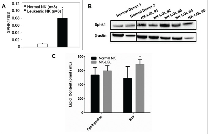
SPHK1 is overexpressed in leukemic NK cells and contributes to a dysregulated sphingolipid rheostat. (A) Quantitative real-time PCR was performed to measure levels of SPHK1 mRNA in PBMC from NK-LGL leukemia patients (CD3−CD56+ >80%, n = 8) or purified NK cells isolated from normal donors (n = 8). SPHK1 mRNA levels are expressed relative to 18S (Mean ± SEM) *, P < 0.05 (Mann-Whitney test). (B) Immunoblot analysis of SPHK1 protein in NK-LGL patient cells or purified NK isolated from normal donors. Loading of protein was confirmed by probing for β-actin. The vertical black line represents a break in the gel where an empty lane was present. (C) Levels of sphingosine and S1P were determined by mass spectrometry in sera from NK-LGL leukemia patients (n = 8) or normal donors (n = 8) (pmol / mL of sera). *, P < 0.05 indicates leukemic cells versus normal NK cells (Student's t-test).