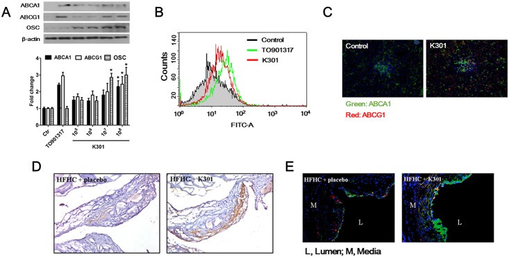
Fig 6. L. acidophilus K301 increased gene expression of ABCA1, ABCG1, and OSC in mouse primary macrophages.
(A) Mouse peritoneal macrophages isolated from ApoE-/- mice were incubated with heat-killed L. acidophilus K301 for 24 h and the expression of ABCA1, ABCG1, and OSC was examined by western blotting using specific antibodies. The amount of protein expression was normalized to that of β-actin. Densitometry analysis was performed by ImageJ software after repeated experiments (lower panel). The error bars in individual samples are shown as variations from triplicate assays. (B) ApoE-/- mice were intraperitoneally injected with 1×10¹¹/kg heat-killed L. acidophilus K301 or TO901317. At 48 h after injection, macrophages were isolated and the expression of ABCA1 was examined by flow cytometric analysis. (C) The expression of ABCA1 and ABCG1 in macrophages isolated from heat-killed L. acidophilus K301-injected ApoE-/- mice was visualized by immunofluorescence staining. (D) Representative immunostaining images showing the change in expression of ABCA1 in ApoE-/- mice fed a high-fat high-cholesterol (HFHC) diet or treated with heat-killed L. acidophilus K301. (E) Immunostaining images of SQLE and OSC in HFHC-fed ApoE-/- mice and L. acidophilus K301-infused ApoE-/- mice; typical images from the area inside the plaques in each group are displayed. In all color images, DAPI (DNA) is shown as blue, whereas target proteins are shown as green (OSC) or red (SQLE). Data shown is representative of three independent experiments.

