Figure 7.

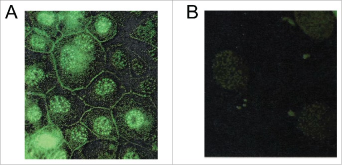
Immunofluorescence detection of Menin localization. (A and B). A Representative example of immunofluorescence staining for Menin in mouse insulinoma cell line TGP61 with the anti-menin antibody (Ab80) (A) or with IgG as a control (B).

Immunofluorescence detection of Menin localization. (A and B). A Representative example of immunofluorescence staining for Menin in mouse insulinoma cell line TGP61 with the anti-menin antibody (Ab80) (A) or with IgG as a control (B).