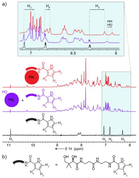
Figure 3.
Pyrrole NMR shift analyses. a, 1H-NMR experiment of pyrrolyl-N-pantetheine probe isolated (red), with apo-PltL (blue), and covalently attached to PltL (green). The enlarged spectra reveal perturbations of pyrrole protons, suggesting pyrrole-PltL interactions. b, Structure of pyrrolyl-N-pantetheine probe.

