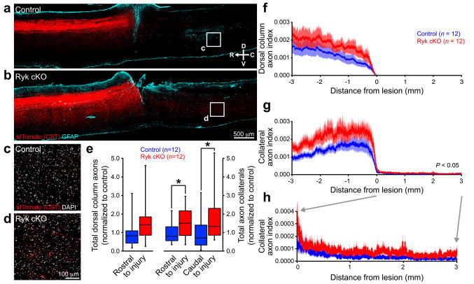
Figure 2. Ryk conditional deletion enhances corticospinal axon sprouting after spinal cord injury.
(a,b) Representative images of tdTomato-labeled CST axons from 8 serial sagittal cryosections spaced 140μm apart superimposed over GFAP astroglial staining at the center of injury (1 experiment, n=12 mice/group, from 11 litters; compass showing dorsal (D), ventral (V), rostral (R), and caudal (C)). Mice with Ryk conditional deletion had greater levels of collateralization both rostral and caudal to the lesion than control mice (one-tailed t-test *P<0.05). (c,d) Higher magnification of single confocal planes from boxed regions (1.5mm caudal to the lesion site) indicated in (a) and (b), respectively. (e) Sum of tdTomato labeled axons (normalized to pyramidal labeling) over 3mm rostral to lesion relative to control in the dorsal columns (n=12 mice/group, one-tailed t-test P=0.12, t(21)=1.198) and in the gray matter. Mice with Ryk conditional deletion had greater levels of collateralization both rostral and caudal to the lesion than control mice (n=12 mice/group, one-tailed t-test *P<0.05: rostral P=0.0499 t(19)=1.730, caudal P=0.0397 t(19)=1.855). (f–h) Distribution of corticospinal axons (axon index is thresholded pixels at every 0.411μm in 8 total saggital spinal cord cryosections divided by thresholded pixels in transverse pyramids) within the dorsal columns (f) or spinal gray matter (g,h). (h) Magnified view of rostral collaterals from (g). C5 injury site is at 0 μm, rostral is represented with negative numbers, caudal with positive. Data in (e) presented as median with inter-quartile range, data in (f–h) presented as mean±s.e.m.

