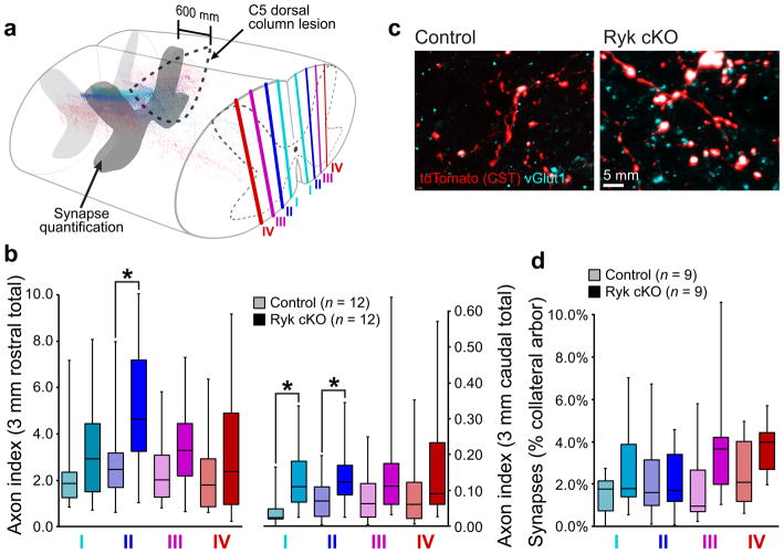
Figure 3. Changes of corticospinal connectivity after C5 dorsal column lesion.
(a–b) Medio-lateral distribution of corticospinal axons shows highest increase in collaterals proximal to the main, dorsal corticospinal tract (regions I and II) after Ryk conditional deletion (n=12 mice/group, one-tailed t-test *P<0.05: II rostral P=0.0225 t(19)=2.146, II caudal P=0.0295 t(21)=1.996, I caudal P=0.0059 t(17)=2.819). (c) Both control and Ryk conditional deletion mice showed pre-synaptic densities (vGlut1 colocalization with tdTomato-labeled corticospinal axons) at 600μm rostral to the C5 injury site (1 experiment, n=9mice/group). (d) Medio-lateral distribution of corticospinal innervation at 600μm rostral to C5 injury site. All data presented as median and inter-quartile range.

