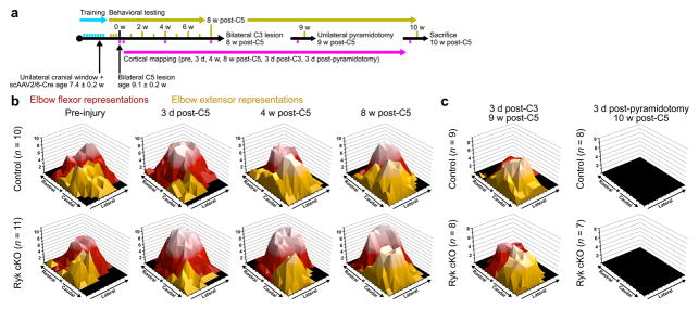
Figure 6. Cortical map re-organization during recovery from spinal cord injury.
(a) Timeline outlining experimental details of optogenetic mapping with weekly behavioral testing following bilateral C5 dorsal column lesion. (b) Topographic representation of elbow flexor and extensor activation, relative to bregma (*) prior to and 3 days, 4 weeks, and 8 weeks after C5 dorsal column lesion. Data presented as total number of mice responding with evoked movements at each location, lighter color indicates a larger number of mice are responsive at a given location. Each tic mark represents 300μm. (c) Subsequent C3 dorsal column lesion disrupts remodeled circuitry, while subsequent pyradmidotomy eliminates unilateral evoked motor output. Both measured at 3 days after injury.

