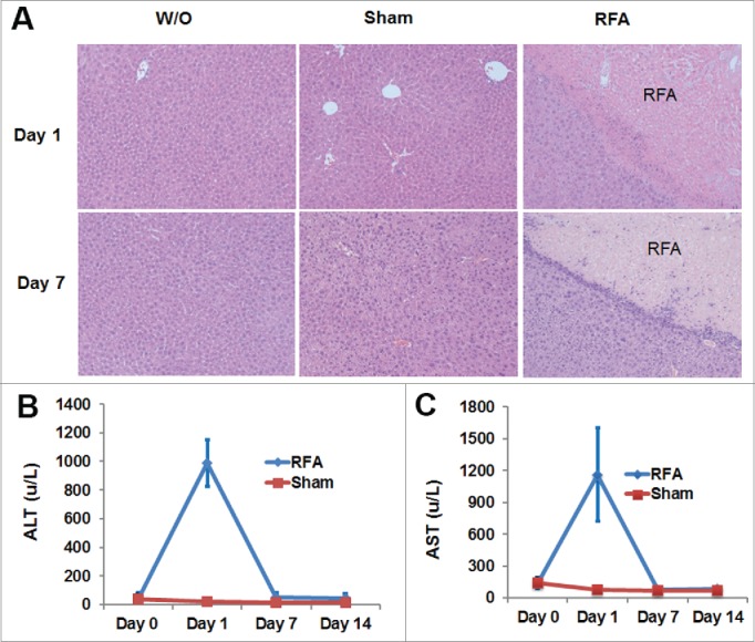
Figure 3.

H& E staining of liver tissue and the levels of ALT and AST in the blood of RFA-treated mice. 10 week-old of C57BL/6 mice received RFA treatment as described. Liver tissue was harvested from each mouse to make slides for H & E staining. Serum was collected for ALT and AST measurements. (A) Representative H & E staining showed RFA-generated liver damage. (B) The level of ALT in the serum from mice with or without RFA treatment. (C) The level of AST in the serum from mice with or without RFA treatment. n=3, *p<0.05, error bars represent mean ±SDs.
