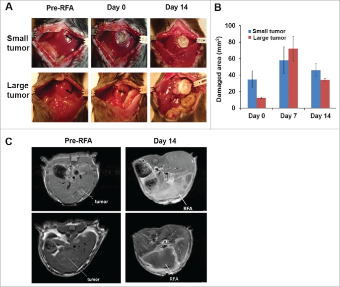
Figure 4.

RFA treatment for destroying small and large tumors. Orthotopic murine HCCs were established, and the tumor size was measured with MRI. Mice with small and large tumors were selected to perform RFA with parameters that were previously defined in normal mice. A power output of 10 W for 60 s was utilized in tumor ablation. The temperature of the probe tip was set at 85°C. (A) Representative macroscopic pictures showed tumor ablation after RFA treatment. (B) Accumulated results demonstrate the damaged liver area post-RFA application. n=3, *p<0.05, error bars represent mean ±SDs. (C) Representative MRI scans to monitor tumor and RFA-generated tumor damage.
