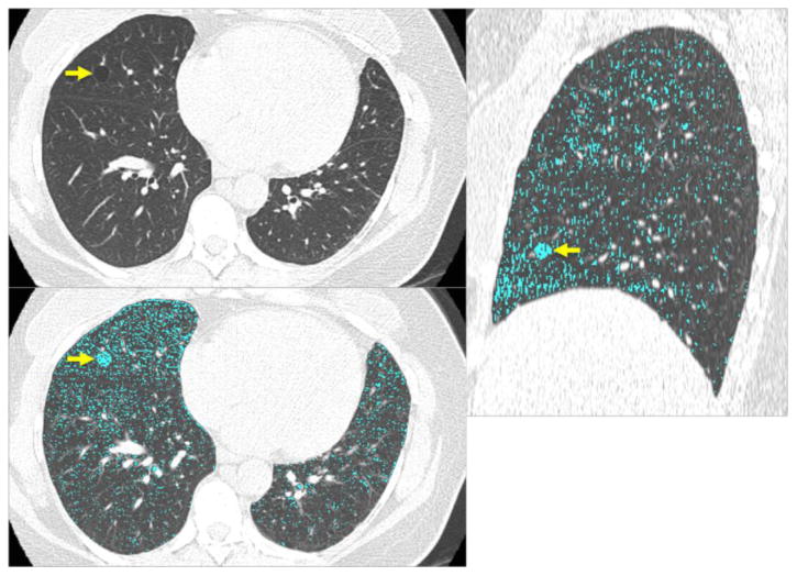
Figure 2.

CT images of a 47-year-old female show a solitary round cyst (arrow) with a thin wall in the middle lobe of the right lung (a). She is a former smoker with 18 pack-years. (b) Low attenuation color-mapping overlaid images (threshold of -950 HU, indicated in light blue areas) at transaxial (b) and sagittal reconstruction (c) demonstrate the distinct solitary pulmonary cyst (arrows) with the background lung parenchyma with a diffuse infiltration of mild centrilobular emphysema.
