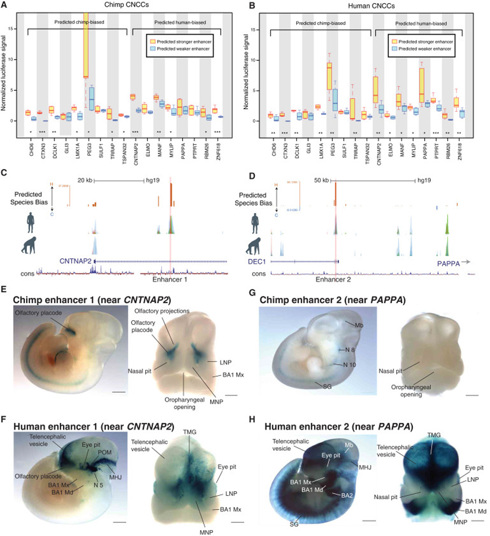
Figure 3. In vitro and in vivo validations of species-biased enhancers.

(A) –(B) Luciferase reporter assays performed in chimp CNCCs (A) or human CNCCs (B) for 9 chimp-biased regions (and orthologous weak human enhancers) and 8 human-biased regions (and orthologous weak chimp enhancers). Luciferase signal was normalized to renilla transfection control. Significance tested from three biological replicates from each species with ANOVA followed by residuals testing with Student’s t-test, p-value indicators *<0.05, **<0.01 ***<0.001
(C)–(D) Genome browser tracks showing human-biased Enhancer 1 (near CNTNAP2 gene; C) and Enhancer 2 (near PAPPA gene; D) selected for a lacZ reporter mouse transgenesis assay.
(E)–(F) Analysis of enhancer activity for chimpanzee and human Enhancer 1 in a lacZ reporter transgenic mouse assay. (E) Representative E11.5 transgenic embryo obtained for the chimpanzee Enhancer 1 reporter, shown in lateral view (left) or frontal view (right) of the embryonic head. (F) Representative E11.5 transgenic embryo obtained for the human Enhancer 1 reporter, shown in lateral view (left) or frontal view (right). Midbrain/hindbrain junction (MHJ); periocular mesenchyme (POM); lateral and medial nasal processes (LNP and MNP); maxillary (Mx) and mandibular (Md) processes of branchial arch 1 (BA1) and BA2. Scale bars: 100 µm (left images) and 50 µm (right images).
(G)–(H) Analysis of enhancer activity for chimpanzee and human Enhancer 2 in a lacZ reporter transgenic mouse assay. (G) Lateral view of representative E11.5 transgenic embryo obtained for the chimpanzee Enhancer 2 reporter, shown in lateral view (left) or frontal view (right) of the embryonic head. (H) Representative E11.5 transgenic embryo obtained for the human Enhancer 2 reporter, shown in lateral view (left) or frontal view (right). Midbrain (Mb); cranial nerves VIII and X (N8 and N10 respectively); sympathetic ganglia (SG) telencephalic midline groove (TMG); midbrain/hindbrain junction (MHJ); maxillary (Mx) and mandibular (Md) processes of branchial arch 1 (BA1) and BA2. Scale bars: 100 µm (left images) and 50 µm (right images).
